|
|
Cystic Fibrosis
Mucovicidosis
General Considerations
- Disease of abnormal exocrine gland function
- Autosomal recessive almost always in Caucasians
- Defect in gene which codes for cystic fibrosis transmembrane conductance regulator (CFTR)
- Major clinical manifestations are pulmonary and pancreatic insufficiency
- Elevated concentration of sodium and chloride in sweat
- Most patients are diagnosed by age 1year
Clinical Findings
- Positive sweat chloride test
- Chronic cough
- Recurrent pulmonary infections
- Higher incidence of asthma and allergy
- Diabetes
- Undescended testicles
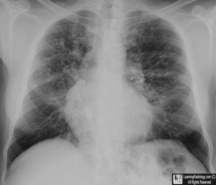
Imaging Findings
- Atelectasis
- Discoid, segmental, lobar with right upper lobe predominance
- Mucoid impaction
- Nodular and fingerlike densities along bronchovascular bundle
- Cylindrical or cystic bronchiectasis
- Hilar adenopathy
- Pulmonary arterial hypertension and cor pulmonale
- Recurrent pneumonias,
- Particularly Staphylococcus, Pseudomonas and P. cepacia
- Clubbing and hypertrophic osteoarthropathy can occur
- Recurrent pneumothorax is common
Differential Diagnosis
- Asthma
- Bronchiectasis
- Aspergillosis
Associated Findings
- Bulky, fatty stools from lack of pancreatic enzymes
- Rectal prolapse
- Meconium ileus — earliest finding
- Meconium ileus equivalent — due to obstruction from stool in older children
- Fatty infiltration of the liver
- Focal biliary cirrhosis with portal hypertension
- Gallstones
- Pancreatic fibrosis due to recurrent Pancreatitis
- Sinusitis
- Hypoplastic frontal sinuses
Treatment
- Goals are to maintain lung function and nutritional therapy
- Bronchodilators
- Chest physical therapy
- Mucolytic agents
- Pancreatic enzyme supplements
- Multivitamins
Prognosis
- Varies from country to country but highest in the United States
- 80% should reach adulthood

Cystic Fibrosis. White arrow points to mucous-filled bronchus; white circles enclose areas of
peribronchial thickening and nodularity. Yellow arrow points to lingular atelectasis.
For this same photo without arrows, click here
For more information, click on the link if you see this icon 
|
|
|
